The human nervous system can be described by both groos anatomy,(which describes the parts that are large enough to be seen with the naked eye,) and by microanatomy, (which describes the system at a cellular level.) In gross anatomy, the nervous system can be divided into two systems: the central nervous system (CNS) and the peripheral nervous system (PNS).[2]
Central Nervous System
See also: List of regions in the human brainThe central nervous system (CNS) is the largest part of the nervous system, and includes the brainand spinal cord. The spinal cavity holds and protects the spinal cord, while the head contains and protects the brain. The CNS is covered by the meninges, a three layered protective coat. The brain is also protected by the skull, and the spinal cord is also protected by the vertebrae.
Peripheral nervous system
The PNS is a regional term for the collective nervous structures that do not lie in the CNS. The bodies of the nerve cells lie in the CNS, either in the brain or the spinal cord, and the longer of the cellular processes of these cells, known as axons, extend through the limbs and the flesh of the torso. The large majority of the axons which are commonly called nerves, are considered to be PNS.
The cell bodies of afferent PNS nerves lie in the dorsal root ganglia.
Microanatomy
The nervous system is, on a small scale, primarily made up of neurons. However, glial cells also play a major role.
Neurons
Neurons are electrically excitable cells in the nervous system that process and transmit information. Neurons are the core components of the brain, the vertebrate spinal cord, the invertebrate ventral nerve cord, and the peripheral nerves. A number of different types of neurons exist: sensory neurons respond to touch, sound, light and numerous other stimuli effecting sensory organs and send signals to the spinal cord and brain, motor neurons receive signals from the brain and spinal cord and cause muscle contractions and affect glands. Interneurons connect neurons to other neurons within the brain and spinal cord.
Glial cells
Glial cells are non-neuronal cells that provide support and nutrition, maintain homeostasis, form myelin, and participate in signal transmission in the nervous system. In the total human brain, the number of glia is estimated to be roughly the same as neurons.[3]
Glial cells provide support and protection for neurons. They are thus known as the "glue" of the nervous system. The four main functions of glial cells are to surround neurons and hold them in place, to supply nutrients and oxygen to neurons, to insulate one neuron from another, and to destroy pathogens and remove dead neurons.
Physiological division
A less anatomical but much more functional way of dividing the human nervous system is classification according to the role that the different neural pathways play, regardless of whether or not they cross through the CNS/PNS:
The somatic nervous system is responsible for coordinating voluntary body movements (i.e. activities that are under conscious control).
The autonomic nervous system is responsible for coordinating involuntary functions, such as breathing and digestion.
In turn, these divisions of the nervous system can be further divided according to the direction in which they conduct nerve impulses:
- Afferent system by sensory neurons, which carries impulses from a somatic receptor to the CNS
- Efferent system by motor neurons, which carries impulses from the CNS to an effector
- Relay system by interneurons (also called "relay neurons"), which transmit impulses between the sensory and motor neurons (both in the CNS and PNS).
The junction between two neurons is called a synapse. There is a very narrow gap (about 20 nm in width) between the neurons called thesynaptic cleft. This is where an action potential (the "message" being carried by the neurons, also known as the nerve impulse) is transmitted from one neuron to the next. This is achieved by relaying the message across the synaptic cleft using neurotransmitters, which diffuse across the gap. The neurotransmitters then bind to receptor sites on the neighboring (postsynaptic) neuron, which in turn produces its own electrical/nerve impulse. This impulse is sent to the next synapse, and the cycle repeats itself.
Nerve impulses are a change in ion balance between the inside and outside of a neuron. Because the nervous system uses a combination of electrical and chemical signals, it is incredibly fast. Although the chemical aspect of signaling is much slower than the electrical aspect, a nerve impulse is still fast enough for the reaction time to be negligible in day to day situations. Speed is a necessary characteristic in order for an organism to quickly identify the presence of danger, and thus avoid injury/death. For example, a hand touching a hot stove. If the nervous system was only comprised of chemical signals, the nervous system would not be able to signal the arm to move fast enough to escape dangerous burns. Thus, the speed of the nervous system is evolutionarily valuable, and is in fact a necessity for life.
Development
Some landmarks of embryonic neural development include the birth and differentiation of neurons from stem cell precursors, the migrationof immature neurons from their birthplaces in the embryo to their final positions, outgrowth of axons from neurons and guidance of the motile growth cone through the embryo towards postsynaptic partners, the generation of synapses between these axons and their postsynaptic partners, and finally the lifelong changes in synapses which are thought to underlie learning and memory.
Importance
The evolution of a complex nervous system makes it possible for various animal species to have advanced perception abilities like sight, complex social interactions, rapid coordination of other organ systems, and integrated processing of many concurrent signals. In humans, the advanced development of the nervous system makes it possible to have language, abstract representation of concepts, transmission of culture, and many other outcomes of human society that would not be possible without our brains.
Many people have lost basic motor skills and other skills because of spinal cord injuries. If this portion is damaged, the biggest nerve and the most important one gets damaged. This leads to paralysis or other permanent damage. Physical lesions or genetic abnormalities of the brain can also lead to major harm.
Abilities
The nervous system enables basic motor skills and sensing. The five classical senses (touch, taste, sight, smell, and hearing) are powered by the nervous system as are others such as equilibrioception (the sensing of gravity), nociception (the sensing of pain), andproprioception (the sensing of relative limb location and motion, as when touching the nose with closed eyes). Inhibition of these senses would retard basic motor skills.
Non-humans
Vertebrates
The nervous system of all vertebrate animals is often divided into the central nervous system (CNS) and the peripheral nervous system (PNS).
Worms
Planaria, a type of flatworm, have dual nerve cords running along the length of the body and merging at the tail and the mouth. These nerve cords are connected by transverse nerves like the rungs of a ladder. These transverse nerves help coordinate the two sides of the animal. Two large ganglia at the head end function similar to a simple brain. Photoreceptors on the animal's eyespots provide sensory information on light and dark.
The nervous system of the roundworm Caenorhabditis elegans has been mapped out to the cellular level. Every neuron and its cellular lineage has been recorded and most, if not all, of the neural connections are known. In this species, the nervous system is sexually dimorphic; the nervous systems of the two sexes, males and hermaphrodites, have different numbers of neurons and groups of neurons that perform sex-specific functions. In C. elegans, males have exactly 383 neurons, while hermaphrodites have exactly 302 neurons [1]
Arthropoda
Arthropods, such as insects and crustaceans, have a nervous system made up of a series of ganglia, connected by a ventral nerve cordmade up of two parallel connectives running along the length of the belly [2]. Typically, each body segment has one ganglion on each side, though some ganglia are fused to form the brain and other large ganglia [3].
The head segment contains the brain, also known as the supraesophageal ganglion. In the insect nervous system, the brain is anatomically divided into the protocerebrum, deutocerebrum, and tritocerebrum. Immediately behind the brain is the subesophageal ganglion, which is composed of three pairs of fused ganglia. It controls the mouthparts, the salivary glands and certain muscles.
Many arthropods have well-developed sensory organs, including compound eyes for vision and antennae for olfaction and pheromonesensation. The sensory information from these organs is processed by the brain.
Development
Neural development in most species has many similarities with neural development in humans.
As shown in a 2008 study, one factor common to all bilateral organisms (including humans) is a familiy of secreted signaling moleculescalled neurotrophins which regulate the growth and survival of neurons[4]. Zhu et al. identified DNT1, the first neurotrophin found in flies. DNT1 shares structural similarity with all known neurotrophins and is a key factor in the fate of neurons in Drosophila. Because neurotrophins have now been identified in both vertebrate and invertebrates, this evidence suggests that neurotrophins were present in an ancestor common to bilateral organisms and may represent a common mechanism for nervous system formation.
The excretory system is a biological system that removes excess, unnecessary or dangerous materials from an organism. It is responsible for the elimination of oxygen waste products of metabolism as well as other nitrogeneous materials. Since the normal operation of most biological systems creates waste, the excretory system is not necessarily distinct from other systems. Instead, it often represents the various excretory processes of several different systems.
Excretory functions
Every organism, from the smallest protist to the largest mammal, must rid itself of the potentially harmful by-products of its own vital activities. This process in living things is called elimination, which may be considered to encompass all of the various mechanisms and processes by which life forms dispose of or throw off waste products, toxic substances, and dead portions of the organism.
Egestion is the act of excreting unusable or undigested material from a cell (as opposed to metabolized waste), as in the case of single-celled organisms, or from the digestive tract of multi-cellular organisms.
As defined above, elimination broadly defines the mechanisms of waste disposal by living systems at all levels of complexity. The term may be used interchangeably with excretion, though not all elimination necessarily takes place in the excretory system.
Component organs
Skin
The skin is another part of the excretory system: it eliminates sweat that helps cool the body and regulate the concentration of salt. The salt helps evaporate the water, cooling off the skin.
Liver
The liver is an organ of the digestive system. It also helps in excreting wastes from the body in a variety of processes. Laboratory analysis reveals a high concentration of a small organelle called a peroxisome, responsible for breakdown of several toxic substances. It also takes in nitrogenous wastes and converts them to urea to reduce their toxicity.
The liver absorbs drugs and other poisonous substances in the blood and changes their chemical structure to make them less toxic and easier to digest. This waste product is called bile and is excreted to the digestive tract, facilitating digestion of fats while also disposing of waste.
Kidneys
The key organs in the excretory system of vertebrates. (See protonephridia system for Platyhelminthes, metanephridia for Annelida, or theMalpighian tubes for insects and terrestrial arthropods.) The kidneys are placed on either side of the spinal column near the lower back. They are primarily responsible for filtering blood by removing nitrogenous wastes, though they also regulate blood pressure in a process called osmoregulation and also assist with the production of red blood cells.
Secretion
The separation, elaboration, and elimination of certain products arising from cellular functions in multi-cellular organisms is called secretion. Though these substances may be a waste product of the cell producing them, they are frequently useful to other cells of the organism. Examples of secretions are the digestive enzymes produced by intestinal and pancreatic tissue cells of vertebrate animals, the hormones synthesized by specialized glandular cells of plants and animals, and sweat secreted by glandular cells in the skins of some humans. Secretion implies that the chemical compounds being secreted were synthesized by specialized cells and that they are of functional value to the cell. The disposal of common waste products should not, therefore, be considered to be of a secretory nature.
The excretory system is a biological system that removes excess, unnecessary or dangerous materials from an organism. It is responsible for the elimination of oxygen waste products of metabolism as well as other nitrogeneous materials. Since the normal operation of most biological systems creates waste, the excretory system is not necessarily distinct from other systems. Instead, it often represents the various excretory processes of several different systems.
[edit]Excretory functions
Every organism, from the smallest protist to the largest mammal, must rid itself of the potentially harmful by-products of its own vital activities. This process in living things is called elimination, which may be considered to encompass all of the various mechanisms and processes by which life forms dispose of or throw off waste products, toxic substances, and dead portions of the organism.
Egestion is the act of excreting unusable or undigested material from a cell (as opposed to metabolized waste), as in the case of single-celled organisms, or from the digestive tract of multi-cellular organisms.
As defined above, elimination broadly defines the mechanisms of waste disposal by living systems at all levels of complexity. The term may be used interchangeably with excretion, though not all elimination necessarily takes place in the excretory system.
[edit]Component organs
[edit]Skin
The skin is another part of the excretory system: it eliminates sweat that helps cool the body and regulate the concentration of salt. The salt helps evaporate the water, cooling off the skin.
[edit]Liver
The liver is an organ of the digestive system. It also helps in excreting wastes from the body in a variety of processes. Laboratory analysis reveals a high concentration of a small organelle called a peroxisome, responsible for breakdown of several toxic substances. It also takes in nitrogenous wastes and converts them to urea to reduce their toxicity.
The liver absorbs drugs and other poisonous substances in the blood and changes their chemical structure to make them less toxic and easier to digest. This waste product is called bile and is excreted to the digestive tract, facilitating digestion of fats while also disposing of waste.
[edit]Kidneys
The key organs in the excretory system of vertebrates. (See protonephridia system for Platyhelminthes, metanephridia for Annelida, or theMalpighian tubes for insects and terrestrial arthropods.) The kidneys are placed on either side of the spinal column near the lower back. They are primarily responsible for filtering blood by removing nitrogenous wastes, though they also regulate blood pressure in a process called osmoregulation and also assist with the production of red blood cells.
[edit]Secretion
The separation, elaboration, and elimination of certain products arising from cellular functions in multi-cellular organisms is called secretion. Though these substances may be a waste product of the cell producing them, they are frequently useful to other cells of the organism. Examples of secretions are the digestive enzymes produced by intestinal and pancreatic tissue cells of vertebrate animals, the hormones synthesized by specialized glandular cells of plants and animals, and sweat secreted by glandular cells in the skins of some humans. Secretion implies that the chemical compounds being secreted were synthesized by specialized cells and that they are of functional value to the cell. The disposal of common waste products should not, therefore, be considered to be of a secretory nature.

Arterial system: canals that carry blood from the heart to the organs.
Posterior auricular: vessel carrying blood to the ear.
Occipital: vessel carrying blood to the head.
External carotid: neck vessel carrying blood to the face.
Internal carotid: neck vessel carrying blood to the brain.
Common carotid (left): vessel carrying blood to the left side of the neck.
Brachio-cephalic: main vessel of the arm.
Left subclavian: vessel carrying blood beneath the left clavicle.
Right coronary artery: vessel feeding blood to the tissues of the right side of the heart.
Thoracic aorta: main artery fo the thorax.
Celiac trunk: vessel carrying blood to the thoracic cavity.
Renal: vessel carrying blood to the kidneys.
Superior mesenteric: vessel carrying blood to the upper part of the abdomen.
Abdominal aorta: principal artery in the abdominal area.
Inferior mesenteric: vessel carrying blood to the lower part of the abdomen.
Common iliac: principal artery of the lower limb of a human being.
Internal iliac: internal branch of the iliac artery.
External iliac: external branch of the iliac artery.
Profunda femoris: vessel carrying blood towards the inside of the thigh.
Peroneal: vessel carrying blood to the lower leg.
Lateral plantar: vessel carrying blood to the side of the sole of the foot.
Dorsalis pedis: vessel carrying blood to the dorsal part fo the foot.
Plantar arch: vessel carrying blood to the instep area of the foot.
Medial plantar: vessel carrying blood to the median part of the sole of the foot.
Anterior tibial: vessel carrying blood to the front part of the lower leg.
Posterior tibial: vessel carrying blood to the back part of the lower leg.
Popliliteal: vessel carrying blood to the back of the foot.
Femoral: vessel carrying blood to the thigh.
Superficial palmar arch: vessel situated just beneath the skin of the parmal arch of the hand.
Ulnar: vessel situated in the area of the ulna.
Common interosseous: vessel situated between the two bones of the forearm.
Gonadal or genital: vessel carrying blood to the genital organs.
Radial: vessel situated in the area of the radius.
Brachial: vessel carrying blood to the arm.
Profunda brachial: vessel carrying blood towards the interior of the arm.
Axillary: vessel carrying blood to the armpit.
Right subclavian: vessel carrying blood beneath the right clavicle.
Right vertebral: vessel situated on the right carrying blood to the vertebrae.
Common carotid (right): vessel carrying blood to the right side of the neck.
Superior thyroid: vessel carrying blood to the thyroid.
Lingual: vessel carrying blood to the tongue.
Facial: vessel carrying blood to the face.
Maxillary: vessel carrying blood to the maxillae.
Superficial temporal: vessel carrying blood to the surface of the skin, in the area of the temples
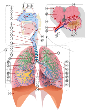
 A complete, schematic view of the human respiratory system.
 A respiratory system's function is to allow gas exchange. The space between the alveoli and the capillaries, the anatomy or structure of the exchange system, and the precise physiological uses of the exchanged gases vary depending on the organism. In humans and other mammals, for example, the anatomical features of the respiratory system include airways, lungs, and the respiratory muscles. Molecules of oxygen and carbon dioxide are passively exchanged, by diffusion, between the gaseous external environment and the blood. This exchange process occurs in the alveolar region of the lungs. [1]
Other animals, such as insects, have respiratory systems with very simple anatomical features, and in amphibians even the skin plays a vital role in gas exchange. Plants also have respiratory systems but the directionality of gas exchange can be opposite to that in animals. The respiratory system in plants also includes anatomical features such as holes on the undersides of leaves known as stomata.
Anatomy of respiratory system in vertebrates
Mammals
For mammals, including humans, respiration is essential. In these organisms, the respiratory system can be subdivided into an upper respiratory tract and a lower respiratory tract based on anatomical features. The upper respiratory tract includes the nasal passages, pharynx and the larynx, while the lower respiratory tract is comprised of the trachea, the primary bronchi and lungs. The respiratory system can also be divided into physiological, or functional, zones. These include the conducting zone (the region for gas transport from the outside atmosphere to just above the alveoli), the transitional zone, and the respiratory zone (the alveolar region where gas exchange occurs). (See also respiratory tract.)
Comparative anatomy/physiology in mammals
Horses
Horses are obligate nasal breathers. That is, they are different from many other mammals in that they do not have the option of breathing through their mouths and must take in air through their nose.
Elephants
The elephant is the only mammal known to have no pleural space. Rather, the parietal and visceral pleura are both composed of dense connective tissue and joined to each other via loose connective tissue. [2] This lack of a pleural space, along with an unusually thick diaphragm, are thought to be evolutionary adaptations allowing the elephant to remain underwater for long periods of time while breathing through its trunk which emerges as a snorkel. [3]
Marine mammals
Rodents
Birds
The respiratory system of birds differs significantly from that found in mammals, containing unique anatomical features such as air sacs. The lungs of birds also do not have the capacity to inflate as birds lack a diaphragm and a pleural cavity. Gas exchange in birds occurs between air capillaries and blood capillaries, rather than in alveoli. See Avian respiratory system for a detailed description of these and other features.
Reptiles
The anatomical structure of the lungs is less complex in reptiles than in mammals, with reptiles lacking the extensive airway tree structure found in mammalian lungs. Gas exchange in reptiles still occurs in alveoli, however. Reptiles do not possess a diaphragm. Thus, breathing occurs via a change in the volume of the body cavity which is controlled by contraction of intercostal muscles in all reptiles except turtles. In turtles, contraction of specific pairs of flank muscles governs inspiration or expiration. [4]
See also reptiles for more detailed descriptions of the respiratory system in these animals.
Amphibians
The skin is one of the important respiratory organs in amphibians. It is highly vascularized and moist, with moisture maintained via secretion of mucus from specialized cells. These properties aid rapid gas exchange.
Fish
In most fish the respiration takes place through gills. (See also aquatic respiration.) Lungfish, however, do possess one or two lungs. The labyrinth fishes have developed a special organ that allows them to take advantage of the oxygen of the air, but is not a true lung.The rare breath fish also has two lungs.
Anatomy of respiratory system in invertebrates
Sponges and jellyfish
These animals lack specialized organs for gas exchange, instead taking in gases directly from the surrounding water.
Flatworms and annelids
Flatworms have special muscles, called "enmmustullus", meaning "small muscles" in Latin. These muscles help the worms to create energy efficiently, while still completing essential activities like eating and sleeping.
Insects
Air enters the respiratory system of most insects through a series of external openings called spiracles. These external openings, which act as muscular valves in some insects, lead to the internal respiratory system, a densely-networked array of tubes called trachea. The tracheal system within an individual is composed of interconnecting transverse and longitudinal tracheae which maintain equivalent pressure throughout the system. These tracheae branch repeatedly, eventually forming tracheoles, which are blind-ended, water-filled compartments only one micrometer in diameter [1]. It is at this level of the tracheoles that oxygen is delivered to the cells for respiration.
Insects were once believed to exchange gases with the environment continuously by the simple diffusion of gases into the tracheal system. More recently, however, large variation in insect ventilatory patterns have been documented and insect respiration appears to be highly variable. Some small insects do demonstrate continuous respiration and may lack muscular control of the spiracles. Others, however, utilize muscular contraction of the abdomen along with coordinated spiracle contraction and relaxation to generate cyclical gas exchange patterns. The most extreme form of these patterns is termed discontinuous gas exchange cycles (DGC) [5].
Molluscs
Molluscs generally possess gills that allow exchange of oxygen from an aqueous envinronment into the circulatory system. These animals also possess a heart that pumps blood which contains hemocyanin as its oxygen-capturing molecule. Hence, this respiratory system is similar to that of vertebrate fish.
Physiology of respiratory system in mammals
For more detailed descriptions see also Respiratory physiology or Respiration.
Ventilation
Ventilation of the lungs is carried out by the muscles of respiration.
Control
Ventilation occurs under the control of the autonomic nervous system from parts of the brain stem, the medulla oblongata and the pons. This area of the brain forms the respiration regulatory center, a series of interconnected brain cells within the lower and middle brain stem which coordinate respiratory movements. The sections are the pneumotaxic center, the apneustic center, and the dorsal and ventral respiratory groups. This section is especially sensitive during infancy, and the neurons can be destroyed if the infant is dropped and/or shaken violently. The result can be death due to "shaken baby syndrome."[6]
Inhalation
Inhalation is initiated by the diaphragm and supported by the external intercostal muscles. Normal resting respirations are 10 to 18 breaths per minute, with a time period of 2 seconds. During vigorous inhalation (at rates exceeding 35 breaths per minute), or in approaching respiratory failure, accessory muscles of respiration are recruited for support. These consist of sternocleidomastoid, platysma, and the scalene muscles of the neck.
Under normal conditions, the diaphragm is the primary driver of inhalation. When the diaphragm contracts, the ribcage expands and the contents of the abdomen are moved downward. This results in a larger thoracic volume and negative (suction) pressure (with respect to atmospheric pressure) inside the thorax. As the pressure in the chest falls, air moves into the conducting zone. Here, the air is filtered, warmed, and humidified as it flows to the lungs.
During forced inhalation, as when taking a deep breath, the external intercostal muscles and accessory muscles aid in further expanding the thoracic cavity.
Exhalation
Exhalation is generally a passive process; however, active or forced exhalation is achieved by the abdominal and the internal intercostal muscles. During this process air is forced or exhaled out.
The lungs have a natural elasticity: as they recoil from the stretch of inhalation, air flows back out until the pressures in the chest and the atmosphere reach equilibrium.[7]
During forced exhalation, as when blowing out a candle, expiratory muscles including the abdominal muscles and internal intercostal muscles, generate abdominal and thoracic pressure, which forces air out of the lungs.
Circulation
The right side of the heart pumps blood from the right ventricle through the pulmonary semilunar valve into the pulmonary trunk. The trunk branches into right and left pulmonary arteries to the pulmonary blood vessels. The vessels generally accompany the airways and also undergo numerous branchings. Once the gas exchange process is complete in the pulmonary capillaries, blood is returned to the left side of the heart through four pulmonary veins, two from each side. The pulmonary circulation has a very low resistance, due to the short distance within the lungs, compared to the systemic circulation, and for this reason, all the pressures within the pulmonary blood vessels are normally low as compared to the pressure of the systemic circulation loop.
Gas exchange
The major function of the respiratory system is gas exchange between the external environment and an organism's circulatory system. In humans and mammals, this exchange facilitates oxygenation of the blood with a concomitant removal of carbon dioxide and other gaseous metabolic wastes from the circulation. As gas exchange occurs, the acid-base balance of the body is maintained as part of homeostasis. If proper ventilation is not maintained, two opposing conditions could occur: 1) respiratory acidosis, a life threatening condition, and 2) respiratory alkalosis.
Upon inhalation, gas exchange occurs at the alveoli, the tiny sacs which are the basic functional component of the lungs. The alveolar walls are extremely thin (approx. 0.2 micrometres). These walls are composed of a single layer of epithelial cells (type I and type II epithelial cells) in close proximity to the pulmonary capillaries which are composed of a single layer of endothelial cells. The close proximity of these two cell types allows permeability to gases and, hence, gas exchange.
Non-respiratory functions
Vocalization
The movement of gas through the larynx, pharynx and mouth allows humans to speak, or phonate. Vocalization, or singing, in birds occurs via the syrinx, an organ located at the base of the trachea. The vibration of air flowing across the larynx (vocal chords), in humans, and the syrinx, in birds, results in sound. Because of this, gas movement is extremely vital for communication purposes.
Temperature control
Panting in dogs and some other animals provides a means of controlling body temperature. This physiological response is used as a cooling mechanism.
Coughing and sneezing
Irritation of nerves within the nasal passages or airways, can induce coughing and sneezing. These responses cause air to be expelled forcefully from the trachea or nose, respectively. In this manner, irritants caught in the mucus which lines the respiratory tract are expelled or moved to the mouth where they can be swallowed.
Development of respiratory system in animals
Humans and mammals
The respiratory system lies dormant in the human fetus during pregnancy. At birth, the respiratory system becomes fully functional upon exposure to air, although some lung development and growth continues throughout childhood. Pre-term birth can lead to infants with under-developed lungs. These lungs show incomplete development of the alveolar type II cells, cells that produce surfactant. The lungs of pre-term infants may not function well because the lack of surfactant leads to increased surface tension within the alveoli. Thus, many alveoli collapse such that no gas exchange can occur within some or most regions of an infant's lungs, a condition termed respiratory distress syndrome. Basic scientific experiments, carried out using cells from chicken lungs, support the potential for using steroids as a means of furthering development of type II alveolar cells.[8] In fact, once a pre-mature birth is threatened, every effort is made to delay the birth, and a series of steroid shots is frequently administered to the mother during this delay in an effort to promote lung growth.[9]
Disease and the respiratory system
Disorders of the respiratory system can be classified into four general areas:
- Obstructive conditions (e.g., emphysema, bronchitis, asthma attacks)
- Restrictive conditions (e.g., fibrosis, sarcoidosis, alveolar damage, pleural effusion)
- Vascular diseases (e.g., pulmonary edema, pulmonary embolism, pulmonary hypertension)
- Infectious, environmental and other "diseases" (e.g., pneumonia, tuberculosis, asbestosis, particulate pollutants): Coughing is of major importance, as it is the body's main method to remove dust, mucus, saliva, and other debris from the lungs. Inability to cough can lead to infection. Deep breathing exercises may help keep finer structures of the lungs clear from particulate matter, etc.
The respiratory tract is constantly exposed to microbes due to the extensive surface area, which is why the respiratory system includes many mechanisms to defend itself and prevent pathogens from entering the body.
Disorders of the respiratory system are usually treated internally by a pulmonologist or respiratory physician1.
Respiratory system in plants
Gas exchange in plants
Plants use carbon dioxide gas in the process of photosynthesis, and then exhale oxygen gas, a waste product of photosynthesis. However, plants also sometimes respire as humans do, taking in oxygen and producing carbon dioxide.
Plant respiration is limited by the process of diffusion. Plants take in carbon dioxide through holes on the undersides of their leaves known as stomata (sing:stoma). However, most plants require little air.[citation needed] Most plants have relatively few living cells outside of their surface because air (which is required for metabolic content) can penetrate only skin deep. However, most plants are not involved in highly aerobic activities, and thus have no need of these living cells.
The human nervous system can be described by both groos anatomy,(which describes the parts that are large enough to be seen with the naked eye,) and by microanatomy, (which describes the system at a cellular level.) In gross anatomy, the nervous system can be divided into two systems: the central nervous system (CNS) and the peripheral nervous system (PNS).[2]
Central Nervous System
The central nervous system (CNS) is the largest part of the nervous system, and includes the brainand spinal cord. The spinal cavity holds and protects the spinal cord, while the head contains and protects the brain. The CNS is covered by the meninges, a three layered protective coat. The brain is also protected by the skull, and the spinal cord is also protected by the vertebrae.
Peripheral nervous system
The PNS is a regional term for the collective nervous structures that do not lie in the CNS. The bodies of the nerve cells lie in the CNS, either in the brain or the spinal cord, and the longer of the cellular processes of these cells, known as axons, extend through the limbs and the flesh of the torso. The large majority of the axons which are commonly called nerves, are considered to be PNS.
The cell bodies of afferent PNS nerves lie in the dorsal root ganglia.
Microanatomy
The nervous system is, on a small scale, primarily made up of neurons. However, glial cells also play a major role.
Neurons
Neurons are electrically excitable cells in the nervous system that process and transmit information. Neurons are the core components of the brain, the vertebrate spinal cord, the invertebrate ventral nerve cord, and the peripheral nerves. A number of different types of neurons exist: sensory neurons respond to touch, sound, light and numerous other stimuli effecting sensory organs and send signals to the spinal cord and brain, motor neurons receive signals from the brain and spinal cord and cause muscle contractions and affect glands. Interneurons connect neurons to other neurons within the brain and spinal cord.
Glial cells
Glial cells are non-neuronal cells that provide support and nutrition, maintain homeostasis, form myelin, and participate in signal transmission in the nervous system. In the total human brain, the number of glia is estimated to be roughly the same as neurons.[3]
Glial cells provide support and protection for neurons. They are thus known as the "glue" of the nervous system. The four main functions of glial cells are to surround neurons and hold them in place, to supply nutrients and oxygen to neurons, to insulate one neuron from another, and to destroy pathogens and remove dead neurons.
Physiological division

A less anatomical but much more functional way of dividing the human nervous system is classification according to the role that the different neural pathways play, regardless of whether or not they cross through the CNS/PNS:
The somatic nervous system is responsible for coordinating voluntary body movements (i.e. activities that are under conscious control).
The autonomic nervous system is responsible for coordinating involuntary functions, such as breathing and digestion.
In turn, these divisions of the nervous system can be further divided according to the direction in which they conduct nerve impulses:
- Afferent system by sensory neurons, which carries impulses from a somatic receptor to the CNS
- Efferent system by motor neurons, which carries impulses from the CNS to an effector
- Relay system by interneurons (also called "relay neurons"), which transmit impulses between the sensory and motor neurons (both in the CNS and PNS).
The junction between two neurons is called a synapse. There is a very narrow gap (about 20 nm in width) between the neurons called thesynaptic cleft. This is where an action potential (the "message" being carried by the neurons, also known as the nerve impulse) is transmitted from one neuron to the next. This is achieved by relaying the message across the synaptic cleft using neurotransmitters, which diffuse across the gap. The neurotransmitters then bind to receptor sites on the neighboring (postsynaptic) neuron, which in turn produces its own electrical/nerve impulse. This impulse is sent to the next synapse, and the cycle repeats itself.
Nerve impulses are a change in ion balance between the inside and outside of a neuron. Because the nervous system uses a combination of electrical and chemical signals, it is incredibly fast. Although the chemical aspect of signaling is much slower than the electrical aspect, a nerve impulse is still fast enough for the reaction time to be negligible in day to day situations. Speed is a necessary characteristic in order for an organism to quickly identify the presence of danger, and thus avoid injury/death. For example, a hand touching a hot stove. If the nervous system was only comprised of chemical signals, the nervous system would not be able to signal the arm to move fast enough to escape dangerous burns. Thus, the speed of the nervous system is evolutionarily valuable, and is in fact a necessity for life.
Development
Some landmarks of embryonic neural development include the birth and differentiation of neurons from stem cell precursors, the migrationof immature neurons from their birthplaces in the embryo to their final positions, outgrowth of axons from neurons and guidance of the motile growth cone through the embryo towards postsynaptic partners, the generation of synapses between these axons and their postsynaptic partners, and finally the lifelong changes in synapses which are thought to underlie learning and memory.
Importance
The evolution of a complex nervous system makes it possible for various animal species to have advanced perception abilities like sight, complex social interactions, rapid coordination of other organ systems, and integrated processing of many concurrent signals. In humans, the advanced development of the nervous system makes it possible to have language, abstract representation of concepts, transmission of culture, and many other outcomes of human society that would not be possible without our brains.
Many people have lost basic motor skills and other skills because of spinal cord injuries. If this portion is damaged, the biggest nerve and the most important one gets damaged. This leads to paralysis or other permanent damage. Physical lesions or genetic abnormalities of the brain can also lead to major harm.
Abilities
The nervous system enables basic motor skills and sensing. The five classical senses (touch, taste, sight, smell, and hearing) are powered by the nervous system as are others such as equilibrioception (the sensing of gravity), nociception (the sensing of pain), andproprioception (the sensing of relative limb location and motion, as when touching the nose with closed eyes). Inhibition of these senses would retard basic motor skills.
Non-humans
Vertebrates
The nervous system of all vertebrate animals is often divided into the central nervous system (CNS) and the peripheral nervous system (PNS).
Worms
Planaria, a type of flatworm, have dual nerve cords running along the length of the body and merging at the tail and the mouth. These nerve cords are connected by transverse nerves like the rungs of a ladder. These transverse nerves help coordinate the two sides of the animal. Two large ganglia at the head end function similar to a simple brain. Photoreceptors on the animal's eyespots provide sensory information on light and dark.
The nervous system of the roundworm Caenorhabditis elegans has been mapped out to the cellular level. Every neuron and its cellular lineage has been recorded and most, if not all, of the neural connections are known. In this species, the nervous system is sexually dimorphic; the nervous systems of the two sexes, males and hermaphrodites, have different numbers of neurons and groups of neurons that perform sex-specific functions. In C. elegans, males have exactly 383 neurons, while hermaphrodites have exactly 302 neurons [1]
Arthropoda
Arthropods, such as insects and crustaceans, have a nervous system made up of a series of ganglia, connected by a ventral nerve cordmade up of two parallel connectives running along the length of the belly [2]. Typically, each body segment has one ganglion on each side, though some ganglia are fused to form the brain and other large ganglia [3].
The head segment contains the brain, also known as the supraesophageal ganglion. In the insect nervous system, the brain is anatomically divided into the protocerebrum, deutocerebrum, and tritocerebrum. Immediately behind the brain is the subesophageal ganglion, which is composed of three pairs of fused ganglia. It controls the mouthparts, the salivary glands and certain muscles.
Many arthropods have well-developed sensory organs, including compound eyes for vision and antennae for olfaction and pheromonesensation. The sensory information from these organs is processed by the brain.
Development
Neural development in most species has many similarities with neural development in humans.
As shown in a 2008 study, one factor common to all bilateral organisms (including humans) is a familiy of secreted signaling moleculescalled neurotrophins which regulate the growth and survival of neurons[4]. Zhu et al. identified DNT1, the first neurotrophin found in flies. DNT1 shares structural similarity with all known neurotrophins and is a key factor in the fate of neurons in Drosophila. Because neurotrophins have now been identified in both vertebrate and invertebrates, this evidence suggests that neurotrophins were present in an ancestor common to bilateral organisms and may represent a common mechanism for nervous system formation.
The excretory system is a biological system that removes excess, unnecessary or dangerous materials from an organism. It is responsible for the elimination of oxygen waste products of metabolism as well as other nitrogeneous materials. Since the normal operation of most biological systems creates waste, the excretory system is not necessarily distinct from other systems. Instead, it often represents the various excretory processes of several different systems.
Excretory functions
Every organism, from the smallest protist to the largest mammal, must rid itself of the potentially harmful by-products of its own vital activities. This process in living things is called elimination, which may be considered to encompass all of the various mechanisms and processes by which life forms dispose of or throw off waste products, toxic substances, and dead portions of the organism.
Egestion is the act of excreting unusable or undigested material from a cell (as opposed to metabolized waste), as in the case of single-celled organisms, or from the digestive tract of multi-cellular organisms.
As defined above, elimination broadly defines the mechanisms of waste disposal by living systems at all levels of complexity. The term may be used interchangeably with excretion, though not all elimination necessarily takes place in the excretory system.
Component organs
Skin
The skin is another part of the excretory system: it eliminates sweat that helps cool the body and regulate the concentration of salt. The salt helps evaporate the water, cooling off the skin.
Liver
The liver is an organ of the digestive system. It also helps in excreting wastes from the body in a variety of processes. Laboratory analysis reveals a high concentration of a small organelle called a peroxisome, responsible for breakdown of several toxic substances. It also takes in nitrogenous wastes and converts them to urea to reduce their toxicity.
The liver absorbs drugs and other poisonous substances in the blood and changes their chemical structure to make them less toxic and easier to digest. This waste product is called bile and is excreted to the digestive tract, facilitating digestion of fats while also disposing of waste.
Kidneys
The key organs in the excretory system of vertebrates. (See protonephridia system for Platyhelminthes, metanephridia for Annelida, or theMalpighian tubes for insects and terrestrial arthropods.) The kidneys are placed on either side of the spinal column near the lower back. They are primarily responsible for filtering blood by removing nitrogenous wastes, though they also regulate blood pressure in a process called osmoregulation and also assist with the production of red blood cells.
Secretion
The separation, elaboration, and elimination of certain products arising from cellular functions in multi-cellular organisms is called secretion. Though these substances may be a waste product of the cell producing them, they are frequently useful to other cells of the organism. Examples of secretions are the digestive enzymes produced by intestinal and pancreatic tissue cells of vertebrate animals, the hormones synthesized by specialized glandular cells of plants and animals, and sweat secreted by glandular cells in the skins of some humans. Secretion implies that the chemical compounds being secreted were synthesized by specialized cells and that they are of functional value to the cell. The disposal of common waste products should not, therefore, be considered to be of a secretory nature.
The excretory system is a biological system that removes excess, unnecessary or dangerous materials from an organism. It is responsible for the elimination of oxygen waste products of metabolism as well as other nitrogeneous materials. Since the normal operation of most biological systems creates waste, the excretory system is not necessarily distinct from other systems. Instead, it often represents the various excretory processes of several different systems.
[edit]Excretory functions
Every organism, from the smallest protist to the largest mammal, must rid itself of the potentially harmful by-products of its own vital activities. This process in living things is called elimination, which may be considered to encompass all of the various mechanisms and processes by which life forms dispose of or throw off waste products, toxic substances, and dead portions of the organism.
Egestion is the act of excreting unusable or undigested material from a cell (as opposed to metabolized waste), as in the case of single-celled organisms, or from the digestive tract of multi-cellular organisms.
As defined above, elimination broadly defines the mechanisms of waste disposal by living systems at all levels of complexity. The term may be used interchangeably with excretion, though not all elimination necessarily takes place in the excretory system.
[edit]Component organs
[edit]Skin
The skin is another part of the excretory system: it eliminates sweat that helps cool the body and regulate the concentration of salt. The salt helps evaporate the water, cooling off the skin.
[edit]Liver
The liver is an organ of the digestive system. It also helps in excreting wastes from the body in a variety of processes. Laboratory analysis reveals a high concentration of a small organelle called a peroxisome, responsible for breakdown of several toxic substances. It also takes in nitrogenous wastes and converts them to urea to reduce their toxicity.
The liver absorbs drugs and other poisonous substances in the blood and changes their chemical structure to make them less toxic and easier to digest. This waste product is called bile and is excreted to the digestive tract, facilitating digestion of fats while also disposing of waste.
[edit]Kidneys
The key organs in the excretory system of vertebrates. (See protonephridia system for Platyhelminthes, metanephridia for Annelida, or theMalpighian tubes for insects and terrestrial arthropods.) The kidneys are placed on either side of the spinal column near the lower back. They are primarily responsible for filtering blood by removing nitrogenous wastes, though they also regulate blood pressure in a process called osmoregulation and also assist with the production of red blood cells.
[edit]Secretion
The separation, elaboration, and elimination of certain products arising from cellular functions in multi-cellular organisms is called secretion. Though these substances may be a waste product of the cell producing them, they are frequently useful to other cells of the organism. Examples of secretions are the digestive enzymes produced by intestinal and pancreatic tissue cells of vertebrate animals, the hormones synthesized by specialized glandular cells of plants and animals, and sweat secreted by glandular cells in the skins of some humans. Secretion implies that the chemical compounds being secreted were synthesized by specialized cells and that they are of functional value to the cell. The disposal of common waste products should not, therefore, be considered to be of a secretory nature.
Arterial system: canals that carry blood from the heart to the organs. A respiratory system's function is to allow gas exchange. The space between the alveoli and the capillaries, the anatomy or structure of the exchange system, and the precise physiological uses of the exchanged gases vary depending on the organism. In humans and other mammals, for example, the anatomical features of the respiratory system include airways, lungs, and the respiratory muscles. Molecules of oxygen and carbon dioxide are passively exchanged, by diffusion, between the gaseous external environment and the blood. This exchange process occurs in the alveolar region of the lungs. [1] Other animals, such as insects, have respiratory systems with very simple anatomical features, and in amphibians even the skin plays a vital role in gas exchange. Plants also have respiratory systems but the directionality of gas exchange can be opposite to that in animals. The respiratory system in plants also includes anatomical features such as holes on the undersides of leaves known as stomata. For mammals, including humans, respiration is essential. In these organisms, the respiratory system can be subdivided into an upper respiratory tract and a lower respiratory tract based on anatomical features. The upper respiratory tract includes the nasal passages, pharynx and the larynx, while the lower respiratory tract is comprised of the trachea, the primary bronchi and lungs. The respiratory system can also be divided into physiological, or functional, zones. These include the conducting zone (the region for gas transport from the outside atmosphere to just above the alveoli), the transitional zone, and the respiratory zone (the alveolar region where gas exchange occurs). (See also respiratory tract.) Horses are obligate nasal breathers. That is, they are different from many other mammals in that they do not have the option of breathing through their mouths and must take in air through their nose. The elephant is the only mammal known to have no pleural space. Rather, the parietal and visceral pleura are both composed of dense connective tissue and joined to each other via loose connective tissue. [2] This lack of a pleural space, along with an unusually thick diaphragm, are thought to be evolutionary adaptations allowing the elephant to remain underwater for long periods of time while breathing through its trunk which emerges as a snorkel. [3] The respiratory system of birds differs significantly from that found in mammals, containing unique anatomical features such as air sacs. The lungs of birds also do not have the capacity to inflate as birds lack a diaphragm and a pleural cavity. Gas exchange in birds occurs between air capillaries and blood capillaries, rather than in alveoli. See Avian respiratory system for a detailed description of these and other features. The anatomical structure of the lungs is less complex in reptiles than in mammals, with reptiles lacking the extensive airway tree structure found in mammalian lungs. Gas exchange in reptiles still occurs in alveoli, however. Reptiles do not possess a diaphragm. Thus, breathing occurs via a change in the volume of the body cavity which is controlled by contraction of intercostal muscles in all reptiles except turtles. In turtles, contraction of specific pairs of flank muscles governs inspiration or expiration. [4] See also reptiles for more detailed descriptions of the respiratory system in these animals. The skin is one of the important respiratory organs in amphibians. It is highly vascularized and moist, with moisture maintained via secretion of mucus from specialized cells. These properties aid rapid gas exchange. In most fish the respiration takes place through gills. (See also aquatic respiration.) Lungfish, however, do possess one or two lungs. The labyrinth fishes have developed a special organ that allows them to take advantage of the oxygen of the air, but is not a true lung.The rare breath fish also has two lungs. These animals lack specialized organs for gas exchange, instead taking in gases directly from the surrounding water. Flatworms have special muscles, called "enmmustullus", meaning "small muscles" in Latin. These muscles help the worms to create energy efficiently, while still completing essential activities like eating and sleeping. Air enters the respiratory system of most insects through a series of external openings called spiracles. These external openings, which act as muscular valves in some insects, lead to the internal respiratory system, a densely-networked array of tubes called trachea. The tracheal system within an individual is composed of interconnecting transverse and longitudinal tracheae which maintain equivalent pressure throughout the system. These tracheae branch repeatedly, eventually forming tracheoles, which are blind-ended, water-filled compartments only one micrometer in diameter [1]. It is at this level of the tracheoles that oxygen is delivered to the cells for respiration. Insects were once believed to exchange gases with the environment continuously by the simple diffusion of gases into the tracheal system. More recently, however, large variation in insect ventilatory patterns have been documented and insect respiration appears to be highly variable. Some small insects do demonstrate continuous respiration and may lack muscular control of the spiracles. Others, however, utilize muscular contraction of the abdomen along with coordinated spiracle contraction and relaxation to generate cyclical gas exchange patterns. The most extreme form of these patterns is termed discontinuous gas exchange cycles (DGC) [5]. Molluscs generally possess gills that allow exchange of oxygen from an aqueous envinronment into the circulatory system. These animals also possess a heart that pumps blood which contains hemocyanin as its oxygen-capturing molecule. Hence, this respiratory system is similar to that of vertebrate fish. For more detailed descriptions see also Respiratory physiology or Respiration. Ventilation of the lungs is carried out by the muscles of respiration. Ventilation occurs under the control of the autonomic nervous system from parts of the brain stem, the medulla oblongata and the pons. This area of the brain forms the respiration regulatory center, a series of interconnected brain cells within the lower and middle brain stem which coordinate respiratory movements. The sections are the pneumotaxic center, the apneustic center, and the dorsal and ventral respiratory groups. This section is especially sensitive during infancy, and the neurons can be destroyed if the infant is dropped and/or shaken violently. The result can be death due to "shaken baby syndrome."[6] Inhalation is initiated by the diaphragm and supported by the external intercostal muscles. Normal resting respirations are 10 to 18 breaths per minute, with a time period of 2 seconds. During vigorous inhalation (at rates exceeding 35 breaths per minute), or in approaching respiratory failure, accessory muscles of respiration are recruited for support. These consist of sternocleidomastoid, platysma, and the scalene muscles of the neck. Under normal conditions, the diaphragm is the primary driver of inhalation. When the diaphragm contracts, the ribcage expands and the contents of the abdomen are moved downward. This results in a larger thoracic volume and negative (suction) pressure (with respect to atmospheric pressure) inside the thorax. As the pressure in the chest falls, air moves into the conducting zone. Here, the air is filtered, warmed, and humidified as it flows to the lungs. During forced inhalation, as when taking a deep breath, the external intercostal muscles and accessory muscles aid in further expanding the thoracic cavity. Exhalation is generally a passive process; however, active or forced exhalation is achieved by the abdominal and the internal intercostal muscles. During this process air is forced or exhaled out. The lungs have a natural elasticity: as they recoil from the stretch of inhalation, air flows back out until the pressures in the chest and the atmosphere reach equilibrium.[7] During forced exhalation, as when blowing out a candle, expiratory muscles including the abdominal muscles and internal intercostal muscles, generate abdominal and thoracic pressure, which forces air out of the lungs. The right side of the heart pumps blood from the right ventricle through the pulmonary semilunar valve into the pulmonary trunk. The trunk branches into right and left pulmonary arteries to the pulmonary blood vessels. The vessels generally accompany the airways and also undergo numerous branchings. Once the gas exchange process is complete in the pulmonary capillaries, blood is returned to the left side of the heart through four pulmonary veins, two from each side. The pulmonary circulation has a very low resistance, due to the short distance within the lungs, compared to the systemic circulation, and for this reason, all the pressures within the pulmonary blood vessels are normally low as compared to the pressure of the systemic circulation loop. The major function of the respiratory system is gas exchange between the external environment and an organism's circulatory system. In humans and mammals, this exchange facilitates oxygenation of the blood with a concomitant removal of carbon dioxide and other gaseous metabolic wastes from the circulation. As gas exchange occurs, the acid-base balance of the body is maintained as part of homeostasis. If proper ventilation is not maintained, two opposing conditions could occur: 1) respiratory acidosis, a life threatening condition, and 2) respiratory alkalosis. Upon inhalation, gas exchange occurs at the alveoli, the tiny sacs which are the basic functional component of the lungs. The alveolar walls are extremely thin (approx. 0.2 micrometres). These walls are composed of a single layer of epithelial cells (type I and type II epithelial cells) in close proximity to the pulmonary capillaries which are composed of a single layer of endothelial cells. The close proximity of these two cell types allows permeability to gases and, hence, gas exchange. The movement of gas through the larynx, pharynx and mouth allows humans to speak, or phonate. Vocalization, or singing, in birds occurs via the syrinx, an organ located at the base of the trachea. The vibration of air flowing across the larynx (vocal chords), in humans, and the syrinx, in birds, results in sound. Because of this, gas movement is extremely vital for communication purposes. Panting in dogs and some other animals provides a means of controlling body temperature. This physiological response is used as a cooling mechanism. Irritation of nerves within the nasal passages or airways, can induce coughing and sneezing. These responses cause air to be expelled forcefully from the trachea or nose, respectively. In this manner, irritants caught in the mucus which lines the respiratory tract are expelled or moved to the mouth where they can be swallowed. The respiratory system lies dormant in the human fetus during pregnancy. At birth, the respiratory system becomes fully functional upon exposure to air, although some lung development and growth continues throughout childhood. Pre-term birth can lead to infants with under-developed lungs. These lungs show incomplete development of the alveolar type II cells, cells that produce surfactant. The lungs of pre-term infants may not function well because the lack of surfactant leads to increased surface tension within the alveoli. Thus, many alveoli collapse such that no gas exchange can occur within some or most regions of an infant's lungs, a condition termed respiratory distress syndrome. Basic scientific experiments, carried out using cells from chicken lungs, support the potential for using steroids as a means of furthering development of type II alveolar cells.[8] In fact, once a pre-mature birth is threatened, every effort is made to delay the birth, and a series of steroid shots is frequently administered to the mother during this delay in an effort to promote lung growth.[9] Disorders of the respiratory system can be classified into four general areas: The respiratory tract is constantly exposed to microbes due to the extensive surface area, which is why the respiratory system includes many mechanisms to defend itself and prevent pathogens from entering the body. Disorders of the respiratory system are usually treated internally by a pulmonologist or respiratory physician1. Plants use carbon dioxide gas in the process of photosynthesis, and then exhale oxygen gas, a waste product of photosynthesis. However, plants also sometimes respire as humans do, taking in oxygen and producing carbon dioxide. Plant respiration is limited by the process of diffusion. Plants take in carbon dioxide through holes on the undersides of their leaves known as stomata (sing:stoma). However, most plants require little air.[citation needed] Most plants have relatively few living cells outside of their surface because air (which is required for metabolic content) can penetrate only skin deep. However, most plants are not involved in highly aerobic activities, and thus have no need of these living cells.
Posterior auricular: vessel carrying blood to the ear.
Occipital: vessel carrying blood to the head.
External carotid: neck vessel carrying blood to the face.
Internal carotid: neck vessel carrying blood to the brain.
Common carotid (left): vessel carrying blood to the left side of the neck.
Brachio-cephalic: main vessel of the arm.
Left subclavian: vessel carrying blood beneath the left clavicle.
Right coronary artery: vessel feeding blood to the tissues of the right side of the heart.
Thoracic aorta: main artery fo the thorax.
Celiac trunk: vessel carrying blood to the thoracic cavity.
Renal: vessel carrying blood to the kidneys.
Superior mesenteric: vessel carrying blood to the upper part of the abdomen.
Abdominal aorta: principal artery in the abdominal area.
Inferior mesenteric: vessel carrying blood to the lower part of the abdomen.
Common iliac: principal artery of the lower limb of a human being.
Internal iliac: internal branch of the iliac artery.
External iliac: external branch of the iliac artery.
Profunda femoris: vessel carrying blood towards the inside of the thigh.
Peroneal: vessel carrying blood to the lower leg.
Lateral plantar: vessel carrying blood to the side of the sole of the foot.
Dorsalis pedis: vessel carrying blood to the dorsal part fo the foot.
Plantar arch: vessel carrying blood to the instep area of the foot.
Medial plantar: vessel carrying blood to the median part of the sole of the foot.
Anterior tibial: vessel carrying blood to the front part of the lower leg.
Posterior tibial: vessel carrying blood to the back part of the lower leg.
Popliliteal: vessel carrying blood to the back of the foot.
Femoral: vessel carrying blood to the thigh.
Superficial palmar arch: vessel situated just beneath the skin of the parmal arch of the hand.
Ulnar: vessel situated in the area of the ulna.
Common interosseous: vessel situated between the two bones of the forearm.
Gonadal or genital: vessel carrying blood to the genital organs.
Radial: vessel situated in the area of the radius.
Brachial: vessel carrying blood to the arm.
Profunda brachial: vessel carrying blood towards the interior of the arm.
Axillary: vessel carrying blood to the armpit.
Right subclavian: vessel carrying blood beneath the right clavicle.
Right vertebral: vessel situated on the right carrying blood to the vertebrae.
Common carotid (right): vessel carrying blood to the right side of the neck.
Superior thyroid: vessel carrying blood to the thyroid.
Lingual: vessel carrying blood to the tongue.
Facial: vessel carrying blood to the face.
Maxillary: vessel carrying blood to the maxillae.
Superficial temporal: vessel carrying blood to the surface of the skin, in the area of the temples![]()
Anatomy of respiratory system in vertebrates
Mammals
Comparative anatomy/physiology in mammals
Horses
Elephants
Marine mammals
Rodents
Birds
Reptiles
Amphibians
Fish
Anatomy of respiratory system in invertebrates
Sponges and jellyfish
Flatworms and annelids
Insects
Molluscs
Physiology of respiratory system in mammals
Ventilation
Control
Inhalation
Exhalation
Circulation
Gas exchange
Non-respiratory functions
Vocalization
Temperature control
Coughing and sneezing
Development of respiratory system in animals
Humans and mammals
Disease and the respiratory system
Respiratory system in plants
Gas exchange in plants

No comments:
Post a Comment